
MPFL Reconstruction
Fixing recurring kneecap dislocations for permanent stability.
MPFL Reconstruction
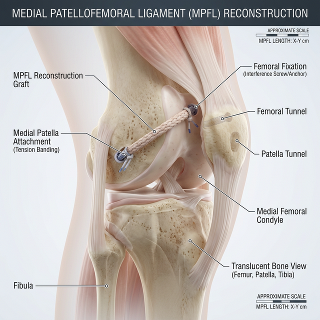
The Medial Patellofemoral Ligament (MPFL) is the primary 'check-rein' that holds the kneecap (patella) in its proper groove. When this ligament is torn—typically during a dislocation—the kneecap becomes unstable and prone to popping out repeatedly. MPFL Reconstruction uses a high-strength graft to replace the torn ligament, restoring the natural tension and track of the patella. Dr. Ravi Teja's advanced technique focuses on precise anatomical anchoring, ensuring the graft isn't too tight (which can cause pain) or too loose (which can lead to failure), resulting in a joint that feels stable and reliable once again.
Surgical Process
Instability Map
Reviewing MRI and CT data to confirm the ligament tear and check for bone malalignment.
Graft Placement
Precisely anchoring a high-strength graft between the femur and the inner edge of the patella.
Tracking Verification
Manually checking the patellar movement throughout a full range of motion before finalizing tension.
Patient Care
Recovery Roadmap
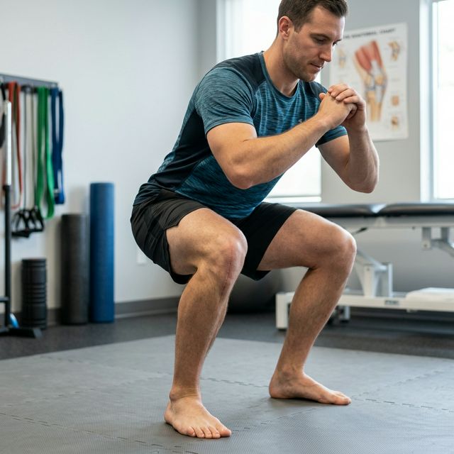
Patients typically use a hinged brace for 6 weeks and can begin early range-of-motion exercises. Full return to sports and high-impact activity is achieved between 4 and 6 months, once quadriceps strength is restored.
Who Needs This Procedure?
Patients with two or more episodes of the kneecap 'popping out' (patella dislocation)
Individuals with a chronic fear of the kneecap sliding out during activity
Patients with a torn Medial Patellofemoral Ligament (MPFL)
Athletes needing stable kneecap tracking to return to high-impact sports
Clinical Results & Gallery
Procedural Intelligence
Elite Patient Care
Dr. Ravi Teja provides personalized evaluations using the latest diagnostic technology for MPFL Reconstruction.
Book EvaluationElite Standard
Certified Excellence
Safety First
Minimal Complications