Cartilage Restoration
Advanced surgical techniques to repair and restore damaged joint surfaces.
Cartilage Restoration
Cartilage fails to heal on its own due to a lack of blood supply. Cartilage Restoration embodies several advanced surgical techniques—such as Microfracture, OATS (Osteochondral Autograft Transfer System), and MACI (Matrix-Induced Autologous Chondrocyte Implantation)—designed to stimulate new cartilage growth or transplant healthy cartilage into a defect. Dr. Ravi Teja meticulously evaluates the size, depth, and location of the 'pothole' to determine the optimal biological strategy, aiming to seamlessly resurface the joint and prevent the onset of early osteoarthritis.
Surgical Process
Lesion Mapping
Using advanced MRI imaging to meticulously measure the cartilage defect.
Biological Preparation
Debriding the damaged area to create a stable, healthy boundary for the graft.
Surface Restoration
Applying the chosen technique (microfracture, plug transfer, or cell scaffold) to precisely fill the defect.
Patient Care
Recovery Roadmap
Recovery is dependent on the specific technique used, but typically involves a period of protected weight-bearing (often 6 weeks) using crutches and a specialized brace. A dedicated physical therapy program is crucial for encouraging the new cartilage to mature and integrate. Return to high-impact sports usually takes 6-12 months.
Who Needs This Procedure?
Active individuals with localized full-thickness cartilage defects (potholes)
Patients with pain localized to one specific area of the knee
Individuals with underlying healthy bone who are too young for joint replacement
Those suffering from osteochondritis dissecans (OCD) lesions
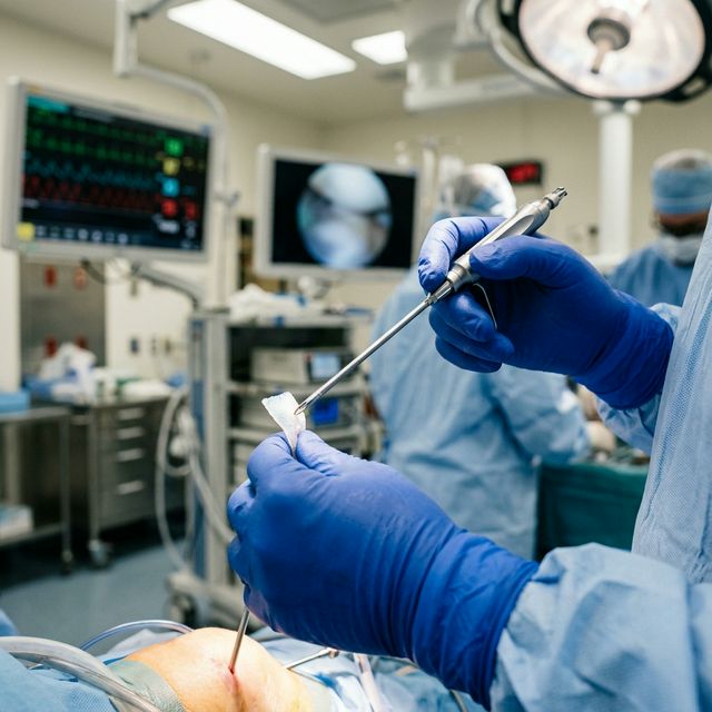
Clinical Results & Gallery
Procedural Intelligence
Elite Patient Care
Dr. Ravi Teja provides personalized evaluations using the latest diagnostic technology for Cartilage Restoration.
Book EvaluationElite Standard
Certified Excellence
Safety First
Minimal Complications